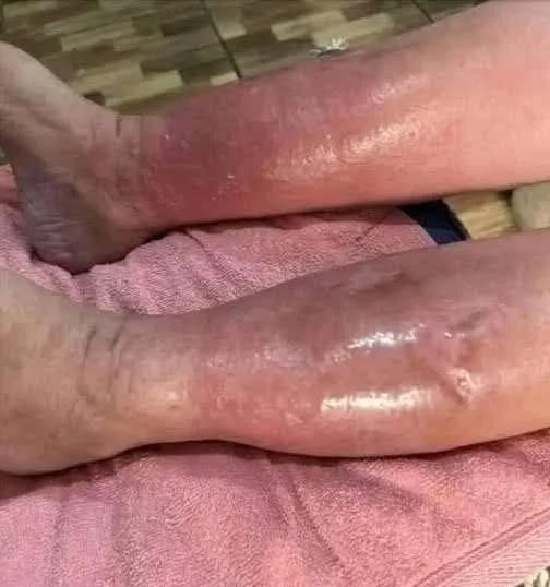
The clinical presentation in image demonstrates severe, diffuse redness (erythema), profound swelling (edema), and a tight, reflective (shiny) appearance of the skin spanning both lower legs. When managing a presentation of this magnitude, the primary medical objective is to differentiate between an infectious cause, a vascular/circulatory insufficiency, or a systemic volume overload.
Potential Etiologies (Causes)
- Venous Insufficiency and Stasis Dermatitis
- Mechanism: This is the most statistically probable cause for bilateral (both legs) involvement. When the valves in the leg veins weaken, blood struggles to return to the heart and pools in the lower extremities.
- Pathology: The resulting high pressure pushes fluid into the surrounding tissues, causing chronic edema. Over time, this fluid irritates the skin from the inside out, leading to severe inflammation, a deep red discoloration, and a shiny texture due to the extreme stretching of the epidermis.
- Bilateral Cellulitis (Acute Bacterial Infection)
- Mechanism: Cellulitis is a deep bacterial skin infection, usually caused by Staphylococcus or Streptococcus species entering through micro-fissures in the skin.
- Pathology: While cellulitis is typically unilateral (affecting only one leg), bilateral presentation can occur, particularly if the skin barrier was already compromised by chronic swelling. It presents as hot, painful, and rapidly spreading redness.
- Systemic Edema (Organ Function Factors)
- Mechanism: When underlying systems—specifically the heart (congestive heart failure), kidneys, or liver—are working inefficiently, the body retains excess fluid.
- Pathology: Gravity causes this fluid to accumulate heavily in the lower legs. If the swelling becomes severe enough, the skin stretches to its absolute limit, triggering a secondary inflammatory response known as stasis erythema.
Medical Management and Treatment Protocols
Because the treatment protocols for these conditions are fundamentally opposite, a definitive in-person physical assessment (including checking for localized warmth, pain response, and peripheral pulses) is mandatory before initiating therapy.
- If Diagnosed as Circulatory (Stasis Dermatitis):
- Compression Therapy: The cornerstone of treatment. Once arterial disease is ruled out, specialized compression wraps or stockings are utilized to force pooled fluid back into circulation.
- Elevation: Patients are instructed to elevate the legs above heart level several times a day to reduce hydrostatic pressure.
- Topical Dermatological Care: High-potency topical corticosteroids may be prescribed briefly to break the inflammatory cycle, alongside heavy emollients to repair the skin barrier.
- If Diagnosed as Infectious (Cellulitis):
- Targeted Antibiotic Therapy: Immediate initiation of systemic oral or intravenous (IV) antibiotics.
- Wound Care: Keeping the skin clean and dry, and marking the borders of the redness to track whether the infection is receding. (Note: Compression wraps are strictly avoided during an active, unmanaged infection).
- If Diagnosed as Systemic Volume Overload:
- Diuretic Therapy: Medications (such as loop diuretics) are prescribed to help the kidneys eliminate excess fluid from the body.
- Underlying Optimization: Direct medical management of the cardiac, renal, or hepatic function.
Critical Red Flags for Immediate Emergency Evaluation
An immediate transition to emergency medical care is required if this presentation is accompanied by:
- A systemic fever, chills, or sudden rigors.
- Rapid upward progression of the redness within a matter of hours.
- The formation of bullae (blisters), open weeping sores, or localized darkening/necrosis of the skin.
- A sudden escalation in localized pain or tenderness.